-
Tutelles et Partenaires
-
Opto-Acoustics
Laser opto-acoustics is a field where acoustic waves, potentially of extremely high frequencies (up to THz), are both generated and detected with short lasers pulses (fs). Recent researches are focused on the dynamic response of single nanoparticles, and on the laser opto-acoustic elastography of single biological cells.

-
- Contact
- Motivation
- Nanoparticle mediated generation of GHz longitudinal acoustic waves
- All-optical probing of a single nanoparticle-substrate contact
- Contact
- Motivation
- Dual opto-acoustic microscopy
- Silica nanoparticles to scatter GHz acoustic phonon
- Remote measurement of a morphological phenotype for cancer cells
- Single cell ultrasonography
- Probing the nano-contact between a cell and a biomaterial
- Speed of sound in a cell organelle with the time resolved Brillouin spectroscopy
- Relaxation dynamics in single polymer microcapsules
- 2019
- 2018
- 2015
- 2014
- 2013
- 2012
Nanophononics
Contact
Motivation
A promising way to locally probe the elasticity of micrometric to nanometric structures (e.g. solid state devices, biological media) is to exploit the high frequency acoustic field radiated by a single nanoparticle (NP). For instance, the femtosecond optical excitation of a 100 nm diameter gold nanoparticle generate in water an acoustic field with a 50 nm acoustic wavelength, i.e. far below the best optical resolution. The aim of this project is to design local GHz opto-acoustic transducers.
Nanoparticle mediated generation of GHz longitudinal acoustic waves
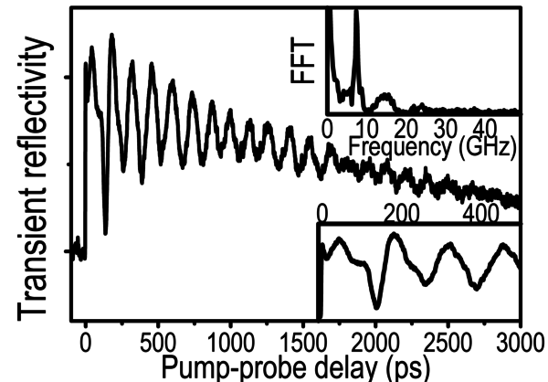
The optoacoustic response of a single submicron (430 nm) gold particle embedded in a silica thin film has been experimentally revealed by transient reflectivity measurements with a femtosecond pump-probe setup. The detection mechanism relies on an intrinsic common-path interferometer where the reference beam comes from a fixed interface of the sample. A semianalytical model is developed to calculate the transient reflectivity accounting for optical index changes in both media and for particle and film surface displacements. The displacement of the particle-film interface turns out to be the major contribution to the measured signal.
Related publications:
Y. Guillet et al. Appl. Phys. Lett. 95, 061909 (2009)
Y. Guillet et al. J. Phys.: Conf. Ser. 214, 012046 (2010)
F. Xu et al. Phys. Rev. B 97, 165412 (2018)
All-optical probing of a single nanoparticle-substrate contact
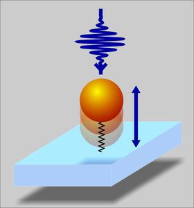
We investigate the GHz dynamics of the elastic contact between a single metallic nanoparticle and a substrate. We detect the known breathing mode of the nanoparticle but we also unravel the axial oscillation of the nanoparticle through an intrinsic common-path interferometer. We measured the eigenfrequency and the lifetime of this vertical motion, which are related to the contact stiffness and hysteresis, and to the acoustic leakage at the nanoparticle-substrate interface. Measurements have been performed for single particles with radii ranging from 60 to 700 nm.
Related publications:
Y. Guillet et al.Phys. Rev. B. 86, 035456 (2012)
Picosecond Biophononics
Contact
Bertrand Audoin & Marie-Fraise Ponge
Motivation
Understanding the mechanical properties of cells and their relation to different physiological conditions is essential to the study of fundamental processes such as proliferation, migration, or differentiation.
The group has pioneered the applications of ultrafast opto-acoustics to probe and image single biological cells. We have demonstrated that the coherent generation of GHz acoustic waves using ultrashort laser pulses, namely picosecond ultrasonics, can efficiently be used to perform single cell ultrasonography. This unconventional cell imaging modality is label free and relies on the organelles mechanical properties as the contrast mechanism.
The technique allows probing the contact between cell and biomaterial with a micron in plane resolution and yields access to the local cell compressibility, remotely. The unequalled capabilities and the foreseen applications suggest tremendous potentialities for single-cell biology.
Dual opto-acoustic microscopy
We used sub-picosecond light pulses to launch high-frequency ultrasound in cells. The dual detection of acoustic echoes and of the time-domain Brillouin scattering allowed us to map remotely and in a single run experiment: the cell adhesion, thickness, storage modulus and mass density, all with a micron resolution. This dual picosecond opto-acoustic microscopy was demonstrated with the multiple imaging of a mitotic macrophage-like cell. The novel modality is compatible with simultaneous standard fluorescence imaging.
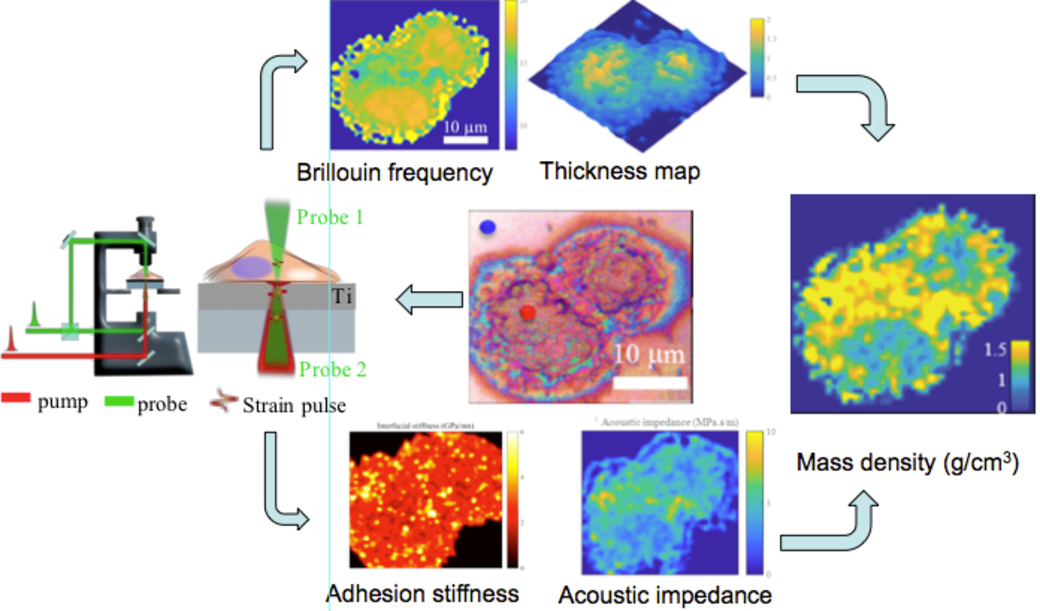 |
Femtosecond lasers for pump (red) and probe (green) were inserted in a standard microscope (left). Acoustic signal from probe 1 allows measurement of the Brillouin frequency shift and allows mapping the cell thickness remotely (top). Acoustic reflectivity measured with probe 2 gives access to the cell adhesion stiffness and to cell acoustic impedance (bottom). By combining data from both arms we mapped the mass density in the nucleus of a mitotic macrophage (right). |
Related publications:
L. Liu et al.Biophotonics 12(8), e201900045 (2019)
Silica nanoparticles to scatter GHz acoustic phonon
When a thin transparent film is immersed in a liquid the optical reflectivity changes may be dominated by the Brillouin response of the liquid, thus hampering detection of the acousto-optic interaction in the sample. We have shown that the undesired signal can be strongly attenuated when silica nanoparticles are sedimented at the sample-liquid interface. The attenuation results from the strong acoustic scattering by the particles of diameter equal to the phonon wavelength. We notably achieved the measurement of the Brillouin frequency shift for a thin film of thickness less than the optical wavelength, without disturbance by the Brillouin interaction in the liquid.
 |
(a) Time domain relative reflectivity changes probed through a thin PMMA film (200 nm) covered with water (blue line) and with silica particles sedimented at the interface between PMMA and water (red line); the magnitudes of signals are normalized with respect to that measured without nanoparticles. Time-frequency wavelet transform of the signal recorded without (b) or with (c) silica particles at the PMMA-water interface. The undesired signal of long duration at 8 GHz vanishes when nanoparticles are sedimented at the sample-water interface. |
Related publications:
M-F Ponge et al.Appl. Phys. Lett. 115, 133101 (2019)
Remote measurement of a morphological phenotype for cancer cells
Cell morphological analysis has long been used in cell biology and physiology for abnormality identification, early cancer detection, and dynamic change analysis under specific environmental stresses. We demonstrated that picosecond ultrasonic microscopy can be used to achieve the remote mapping of cell 3D morphology quantitatively with an in-plane resolution limited by optics and an out-of-plane accuracy down to a tenth of the optical wavelength. For this, we tracked GHz coherent acoustic phonons and their resonance harmonics and mapped the 3D morphology of an entire osteosarcoma cell. The resulting image complies with the image obtained by standard atomic force microscopy, and both revealed very close roughness mean values. In addition, while scanning macrophages and monocytes, we demonstrated an enhanced contrast of thickness mapping by taking advantage of the detection of high-frequency resonance harmonics.
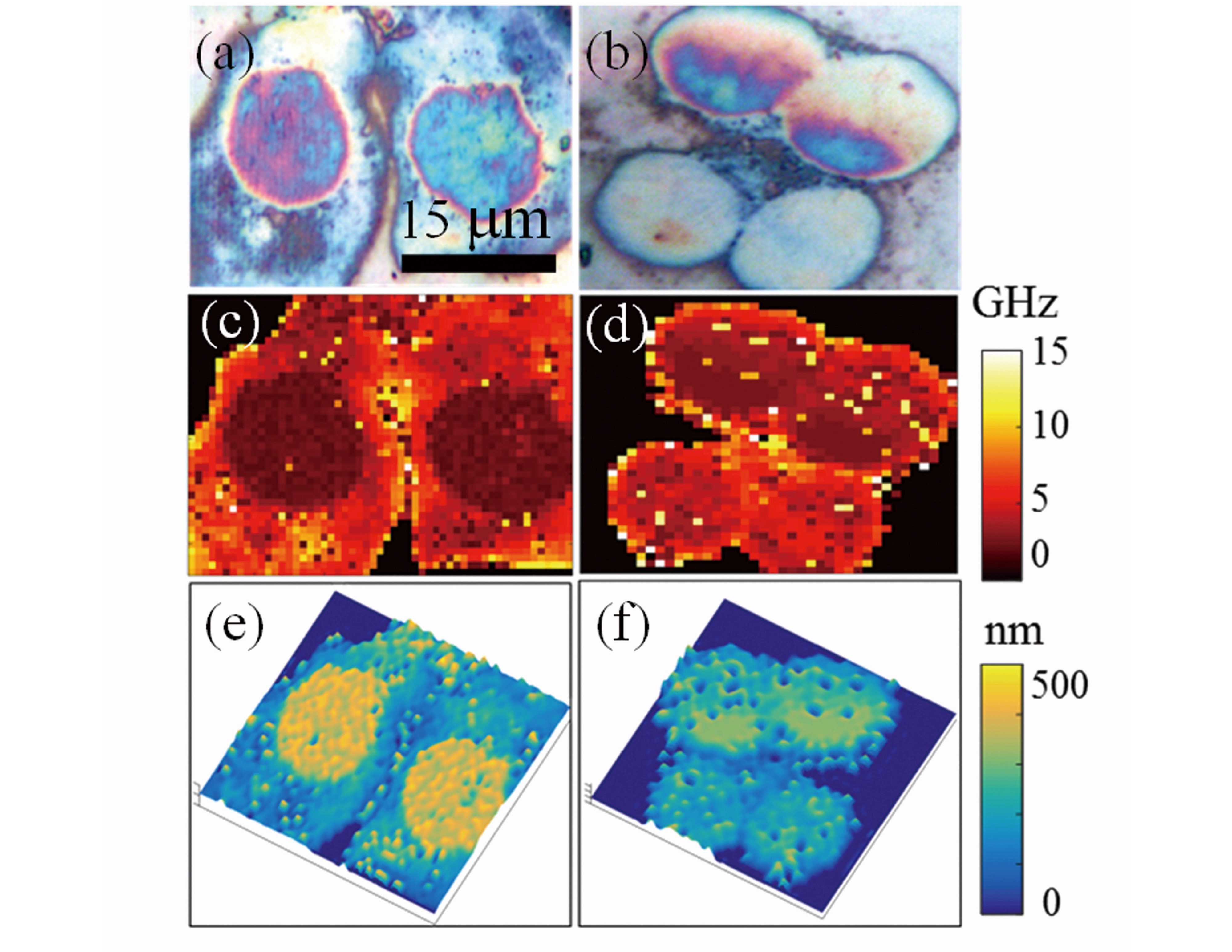 |
Thickness mapping of macrophages (left) and monocytes (right). (a,b) White light top-view images. (c,d) Maps of the acoustic resonances fR measured in the recorded PU signals. (e,f) quantitative 3D cell morphologies. |
Related publications:
L. Liu et al.Scient. Rep. 9, 6409 (2019)
Single cell ultrasonography
We developed an inverted pulsed opto-acoustic microscope (iPOM) operating in the 10 to 100 GHz range. These frequencies allow mapping quantitatively cell structures as thin as 10 nm and resolving the fibrillar details of cells. Using this non-invasive all-optical system, we produce high-resolution images based on mechanical properties as the contrast mechanisms, and we can observe the stiffness and adhesion of single migrating stem cells.
However, manipulating biological media in physiological conditions is often a technical challenge when using a laser-based setup. We have therefore designed a new opto-acoustic bio-transducer composed of a thin metal film sputtered on a transparent heat sink that allows reducing importantly the laser-induced cellular stresses, and offers a wide variety of optical configurations (see figure 1). In particular, by exploiting the acoustic reflection coefficient at the sample-transducer interface and the photoacoustic interaction inside the transparent sample, we have probed simultaneously the density and compressibility of a single vegetal cell.
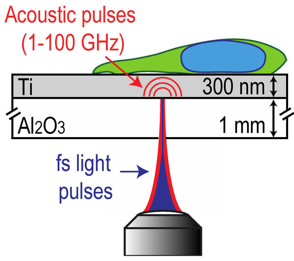 |
Absorption of fs light pulses in a thin metal layer launches longitudinal acoustic pulses with ps dynamics. Coherent phonons propagate through the transducer, are reflected at the metal-cell interface and are optically detected. Scanning laser pulses at the metal interface we image the inhomogeneous reflection of sound by the cell. |
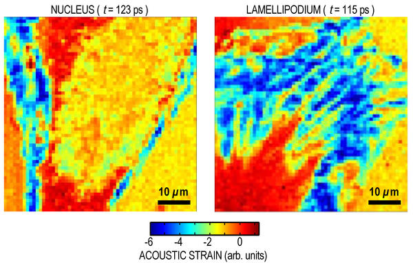 |
Raw acoustic images of the nuclear region and of the lamellipodium of a migrating human mesenchymal stem cell (hMSC) after fixation. The very high acoustic contrast reveals the structure of the nucleus, the actin network and the fine details of the complexity of the adhesion sites at the edge of the lamellipodium. Processing of acoustic signals allows mapping interfacial stiffness and acoustic impedance with same micron resolution. Click on the links to watch nucleus and lamellipodium videoscopy on a ps time frame. |
Related publications:
T. Dehoux et al.Sci. Rep. 5, 8650 (2015)
Probing the nano-contact between a cell and a biomaterial
We have demonstrated the ability of the picosecond ultrasonic technique to image non-specific contacts of single animal cells with metallic substrates. Monocytes were cultured on top of an opto-acoustic bio-transducer. Low-energy femtosecond pump laser pulses were focused at the bottom of the Ti film to a micron spot. The subsequent ultrafast thermal expansion launched a longitudinal acoustic pulse in Ti, with a broad spectrum extending up to 100 GHz. We measured the acoustic echoes reflected from the film/cell interface through the transient optical reflectance changes. The time-frequency analysis of the reflected acoustic pulses with a wavelet transform gives access to a map of the acoustic impedance of the cell and of the stiffness of the film-cell interface.
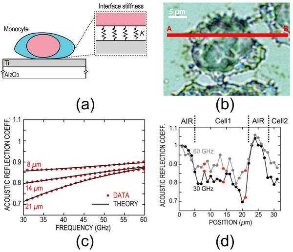 |
a) schematic view of the sample. (b) White-light image of the monocyte and line scanned across the cell. (c) Measured (black plain line) and theoretical (red dashed line) reflection coefficient vs frequency at three positions in the first cell (8, 14 and 20 µm). These positions are indicated in Fig. (d) with red dots. (d) Acoustic reflection coefficient along the cell contact area at frequencies 30 GHz (black) and 60 GHz (grey). |
Related publications:
M. Abi Ghanem et al.J. Biophoton. 7, 453 (2014)
Speed of sound in a cell organelle with the time resolved Brillouin spectroscopy
Absorption of fs laser pulses in the metal launches an acoustic wavefront in the fixed cell. Its propagation along the cell thickness is detected through the Brillouin acousto-optic interaction.[1] The time resolved so-called Brillouin oscillations yield remote measurements of the cell thickness and of the sound velocity and attenuation in the GHz range for vegetal and animal cells.[2]
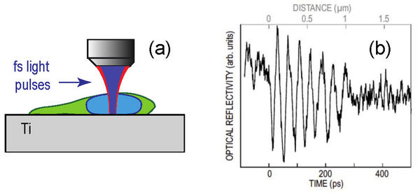 |
(a) Experimental configuration for Brillouin scattering detection. (b) Transient optical reflectivity measured for the intranuclear region of a single osteosarcoma cell. |
We have probed the stiffness and viscosity of nuclei in single animal cells in the previously unexplored GHz range with a 100 nm axial resolution. The probing of cells at contrasted differentiation stages, ranging from stem cells to mature cells originating from different tissues, demonstrates that the mechanical properties of the nuclear network are common across various cell types.[3] This pointed to an asymptotically increasing influence of a solid meshwork of connected chromatin fibres.
We have developed alternative optical configurations. In particular, the acoustic wave was launched at the bottom of a thin metal film and the reflectivity was measured through the cell. By both exploiting the acoustic reflection coefficient at the sample-transducer interface and the photoacoustic interaction inside the transparent sample, the density and compressibility of the sample can be probed simultaneously.[4]
We probed the mechanical properties of vegetal live cells sub-compartiments. Experiments with onion cells revealed that single-cell wall transverse stiffness in the direction perpendicular to the epidermis layer is close to that of cellulose.[5] This observation demonstrated that cellulose micro-fibrils are the main load-bearing structure in this direction, and suggested strong bonding of micro-fibrils by hemicelluloses. Altogether measurement of the viscosity at such high frequencies suggest that the rheology of the wall is dominated by glass-like dynamics, a behaviour attributed to the influence of the pectin matrix.
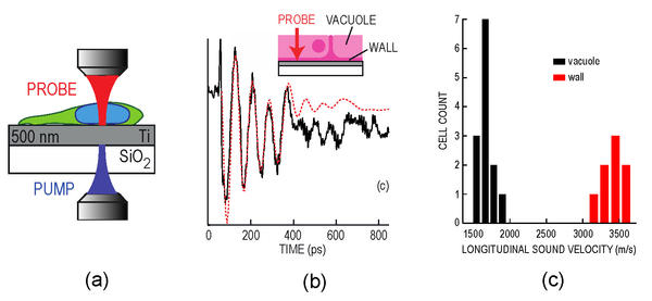 |
(a) Experimental configuration for acoustic reflection coefficient measurements. (b) Transient optical reflectivity measured (plain line) in a thin wall (800 nm) of a vegetal cell. Theoretical calculations are plotted with dotted lines. (c) Longitudinal sound velocity for the cell vacuole (left n = 13) and cell wall (right n = 8). |
Related publications :
[1] C. Rossignol et al., Appl. Phys. Lett.93, 123901 (2008)
[2] M. Ducousso et al., Eur. Phys. J. Appl. Phys. 61, 11201 (2013)
[3] O. F. Zouani et al., Soft Matter 10, 8737-8743 (2014)
[4] T. Dehoux and B. Audoin, J. Appl. Phys 112, 124702 (2012)
[5] A. Gadalla et al., Planta 239, 1129 (2014)
Relaxation dynamics in single polymer microcapsules
Soft polymer micro-objects are of great interest for their biological cell-mimicking properties their encapsulation capacities, their use as contrast agents for ultrasonic imaging, as well as their use in self-healing materials. All the same, their compressibility, viscosity and shell thickness play an important role in the adhesion involved in specific tissues targeting, in the drug release rate, or in their echogenicity. However, at submicrometer scales, controlling these properties is challenging and requires elaborate characterization methods.
In particular, a large body of work has been devoted to microcapsules due to their potential in the food and pharmaceutical industries. Microcapsules are mostly used at MHz frequencies, and their individual rheological behavior is usually inferred from their collective response by modelling. Correlating the input of these models with the mechanics of single capsules at higher frequencies is therefore of the utmost importance to complete the description of their intricate dynamics in view of improving their design and modelling.
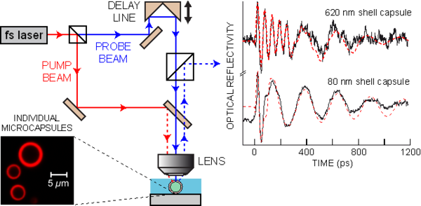
Using an ultrafast optical technique (Fig. x, left) we probe coherent-phonon propagation inside a single microcapsule composed of a nanometric polymer shell made of poly(lactide-co-glycolide) (PLGA) encapsulating a liquid perfluorooctyl bromide (PFOB) core. Longitudinal storage and loss moduli are measured simultaneously in the transparent shell and core at frequencies ~18 and ~4 GHz, respectively, using time-resolved Brillouin spectroscopy (Fig. x, right). A time-frequency analysis allows determination of the thickness of several capsule shells ranging from 620 down to 80 nm. Comparison with lower frequency data shows a weak power-law frequency dependence of phonon attenuation in the PLGA shell, signature of thermally activated processes in glasses.
 |
Related publications :
T. Dehoux et al.Soft Matter 8, 2586 (2012)
Publications
2019
- Scattering of GHz coherent acoustic phonons by silica nanoparticles in water probed with the time-domain Brillouin spectroscopy
M-F Ponge, L. Liu, C. Aristégui and B. Audoin
Appl. Phys. Lett. 115, 133101 (2019) - Label-free multi-parametric imaging of single cells: dual picosecond optoacoustic microscopy
L. Liu, L. Plawinski, M-C Durrieu, and B. Audoin
Biophotonics 12(8), e201900045 (2019) - Remote imaging of single cell 3D morphology with ultrafast coherent phonons and their resonance harmonics
L. Liu, A. Viel, G. Le Saux, L. Plawinski , G. Muggiolu, P. Barberet, M. Pereira, C. Ayela, H. Seznec, M-C Durrieu, J-M Olive and B. Audoin
Scient. Rep. 9, 6409 (2019) - Ultrafast microscopy of the vibrational landscape of a single nanoparticle
Y. Guillet, A. Abbas, S Ravaine. and B. Audoin
Appl. Phys. Lett. 114, 091904 (2019)
2018
- Common-path conoscopic interferometry for enhanced picosecond ultrasound detection
L. Liu, Y. Guillet and B. Audoin
J. Appl. Phys. 123, 173103 (2018) - All-optical in-depth detection of the acoustic wave emitted by a single gold nanorod
F. Xu, Y. Guillet, S. Ravaine and B. Audoin
Phys. Rev. B 97, 165412 (2018) - Directivity of GHz shear acoustic waves launched by the absorption of short laser pulses at the interface between a transparent and an absorbing material
A. Viel, B. Audoin
J. Appl. Phys. 123, 043102 (2018) - Opto-acoustic microscopy reveals adhesion mechanics of single cells
M. Abi Ghanem, T. Dehoux, L. Liu, G. Le Saux, L. Plawinski, MC Durrieu and B. Audoin
Rev. Sci. Instrum. 89, 014901 (2018)
2015
- Thermal microscopy of single biological cells
R. Legrand, M. Abi Ghanem, L. Plawinski, M.-C. Durrieu, B. Audoin and T. Dehoux
Appl. Phys. Lett. 107, 263703 (2015) - Effect of refracted light distribution on the photoelastic generation of ZGV Lamb modes in optically low-absorbing plates
S. Raetz, J. Laurent, T. Dehoux, D. Royer, and C. Prada
J. Acoust. Soc. Am. 138, 3522-3532 (2015) - In-line femtosecond common-path interferometer in reflection mode
J. Chandezon, J.-M. Rampnoux, S. Dilhaire, B. Audoin, and Y. Guillet
Opt. Express 23, 27011 (2015) - Broadband Spectral Signature of the Ultrafast Transient Optical Response of Gold Nanorods
X. L. Wang, Y. Guillet, P. R. Selvakannan, H. Remita, and B. Palpant
J. Phys. Chem. C 119, 7416 (2015) - All-optical broadband ultrasonography of single cells
T. Dehoux, M. Abi Ghanem, O. F. Zouani, J.-M. Rampnoux, Y. Guillet, S. Dilhaire, M.-C. Durrieu and B. Audoin
Sci. Rep. 5, 8650 (2015) - Probing single-cell mechanics with picosecond ultrasonics
T. Dehoux, M. Abi Ghanem, O. F. Zouani, M. Ducousso, N. Chigarev, C. Rossignol, N. Tsapis, M.-C. Durrieu and B. Audoin
Ultrasonics 56, 160-171 (2015)
2014
- Nanoscale mechanical contacts mapped by ultrashort time-scale electron transport
M. Tomoda, T. Dehoux, Y. Iwasaki, O. Matsuda, V. Gusev and O. B. Wright
Sci. Rep. 4, 4790 (2014) - Picosecond time resolved opto-acoustic imaging with 48 MHz frequency resolution
A. Abbas, Y. Guillet, J.-M. Rampnoux, P. Rigail, E. Mottay, B. Audoin and S. Dilhaire
Opt. Express 22, 7831 (2014) - Transverse mechanical properties of cell walls of single living plant cells probed by laser-generated acoustic waves
A. Gadalla, T. Dehoux and B. Audoin
Planta 239, 1129 (2014) - Universality of the network-dynamics of the cell nucleus at high frequencies
O. F. Zouani, T. Dehoux, M.-C. Durrieu and B. Audoin
Soft Matter 10, 8737-8743 (2014) - Remote opto-acoustic probing of single-cell adhesion on metallic surfaces
M. Abi Ghanem, T. Dehoux, O. F. Zouani, A. Gadalla, M.-C. Durrieu and B. Audoin
J. Biophoton. 7, 453 (2014)
2013
- Evaluation of mechanical properties of fixed bone cells with sub-micrometer thickness by picosecond ultrasonics M. Ducousso, O. F. Zouani, C. Chanseau, C. Chollet, C. Rossignol, B. Audoin and M.-C. Durrieu
Eur. Phys. J. Appl. Phys. 61, 11201 (2013) - Acoustic beam steering by light refraction: Illustration with directivity patterns of a tilted volume photoacoustic source
S. Raetz, T. Dehoux, M. Perton and B. Audoin
J. Acoust. Soc. Am. 134, 4381 (2013) - Listening to cells: a non-contact optoacoustic nanoprobe
T. Dehoux, O. F. Zouani, B. Audoin and M.-C. Durrieu
Biophys. J. 104, 193A (2013) - Laser-generated GHz acoustic waves reveal a universal nuclear stiffness probed during cell differentiation
O. F. Zouani, T. Dehoux, M.-C. Durrieu and B. Audoin
Biophys. J. 104, 478A-479A (2013)
2012
- Non-invasive optoacoustic probing of the density and stiffness of single biological cells
T. Dehoux and B. Audoin
J. Appl. Phys 112, 124702 (2012) - All-optical ultrafast spectroscopy of a single nanoparticle-substrate contact
Y. Guillet, B. Audoin, M. Ferrié and S. Ravaine
Phys. Rev. B 86, 035456 (2012) - Scaled behavior of interface waves at an imperfect solid-solid interface
T. Valier-Brasier, T. Dehoux and B. Audoin
J. Appl. Phys. 112, 024904 (2012) - Relaxation dynamics in single polymer microcapsules probed with laser-generated GHz acoustic waves
T. Dehoux, N. Tsapis and B. Audoin
Soft Matter 8, 2586 (2012)
Mise à jour le 22/02/2021
Suivez-nous :

